ICTpost Radiology Bureau
In India more than 400 million people have stress, which is one of the major reasons of all health problems, and change in lifestyles is a major reason causing cardio-vascular diseases. The health profile of the country’s people is becoming more vulnerable to heart diseases, and around 12-14% of the country?s population is likely to suffer from cardiac diseases in their lifetime. According to a study, about 25 per cent of deaths in the age group of 25- 69 years occur because of heart diseases in India. In urban areas, 32.8 % deaths occur because of heart ailments, while 22.9 % people die of heart diseases in rural India. Cardio-vascular diseases in women in India, where a large percentage of the population is diabetic, are likely to increase by 17 percent in the coming 10 years.
In the recent years payments for myocardial perfusion SPECT exceeded $2 billion, rendering it one of the big ticket items in Indian healthcare budget. The reasons for this dramatic utilization of cardiac SPECT imaging can be attributed to a demographic spike of an aging Indian population as well as a sharp uptick in obesity, and concurrent diabetes in those under age 65.
Diabetes has emerged as a major healthcare problem in India. According to Diabetes Atlas published by the International Diabetes Federation (IDF), there were an estimated 40 million persons with diabetes in India in 2007 and this number is predicted to rise to almost 70 million people by 2025. The countries with the largest number of diabetic people will be India, China and USA by 2030. It is estimated that every fifth person with diabetes will be an Indian.
Developers of SPECT equipment and software have kept pace with the modality’s imaging utilization curve. The past several years have seen new developments in both hardware technology and image-processing algorithms that provide substantial reductions in SPECT acquisition time without a sacrifice in diagnostic quality.
At the component level there have been improvements in scintillators and photon transducers as well as a greater availability of semiconductor technology, wrote Mark T. Madsen, PhD, professor and director of the clinical CT research facility at the University of Iowa in the Journal of Nuclear Medicine (April 2007). These devices permit the fabrication of smaller and more compact systems that can be customized for particular applications.
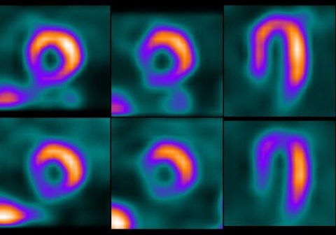
Of particular note has been the development of high-count cardiac SPECT systems that do not use conventional collimation as well as the introduction of hybrid SPECT/CT technology. One high-speed SPECT prototype, utilizing nine pixilated solid-state detector columns, cadmium zinc telluride (CZT) crystals, wide-angle tungsten collimators, region of interest-centric scanning, and increased angular sampling, has shown remarkable benefit for myocardial perfusion imaging (MPI).
The technology has recently been shown to provide an 8- to 10-fold increase in sensitivity, coupled with a two-fold improvement in spatial resolution, and has enabled a significant reduction in imaging time and dose of radioisotopes.
The commercial launch of SPECT/CT systems by Philips Healthcare, GE Healthcare and Siemens Healthcare over the past decade has played a strong role in the growing utilization of SPECT for CAD imaging. These hybrid modalities have allowed the direct fusion of morphologic and functional information for physicians, providing even greater clinical clarity and better patient management.
One of the greatest advantages to using SPECT/CT is that the specificity of the diagnoses has been greatly enhanced. It provides the capability to view the functional and anatomic aspects of the exam together, we are very confident about the localization of what we see. Overall, it has improved tremendously our ability to diagnose accurately.
SPECT/CT has provided another benefit to the facility; by its capability to increase diagnostic certainty, physicians are now better able to provide a diagnosis without conducting additional imaging studies on other modalities. This, in turn, has allowed greater utilization of these technologies for other patients. Clinicians have found tremendous benefit to the attenuation correction available with the CT portion of the hybrid technology.
The CT portion of the SPECT/CT has better imaging characteristics and is not as limiting as the sealed-source method. In addition, sealed sources have to be replaced every 12 to 18 months due to their half-life, which poses an additional problem. It also just doesn’t have the strength of CT alone for the morbidly obese patient. editor@ictpost.com


